

Anatomical Models, Anatomy Charts, Anatomy Posters. We offers human anatomy models, human anatomy chart, educational human anatomy models. Human Anatomical Models, Custom Anatomy Model Manufacturer.
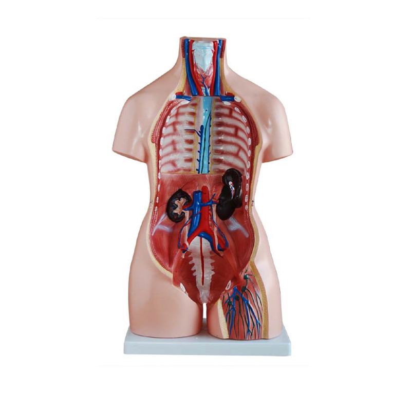
Life size sexless torso showing visceral organs mounted on a base, composed of 8 parts, major organ systems are represented with great attention to accuracy and detail.
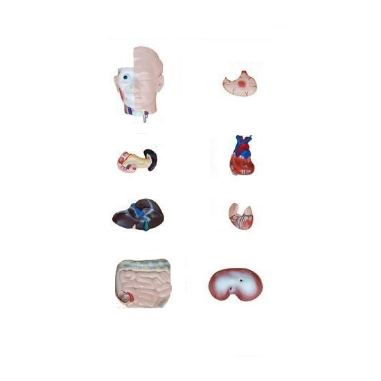
Dissectible into 10 parts, showing ribs, sternum, clavicle with their attachments
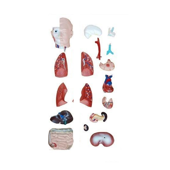
Dissectible into 10 parts, showing ribs, sternum, clavicle with their attachments. Thoracic and abdominal organs detachable individually like lungs,heart, stomach, liver, intestine, half of the skull cap is removable to show brain and its structure, which can be taken out, sectioned head and neck are showing internal organs of that region, natural size, on base with key card.
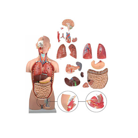
Natural size dissectible into 18 parts, mounted on base with key card. Showing ribs, sternum, clavicle with their attachments, thoracic and abdominal organs detachable individually like lungs, heart, stomach, liver, intestine for detailed study. Half of the skull cap is removable to show brain and its structure, which can be taken out, sectioned head and neck are showing internal organs of that re.
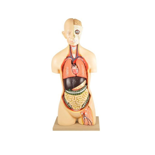
This life size sexless torso mounted on a base, composed of 16 parts, All of the major organ systems are represented with great attention to accuracy and detail. Structures are numbered and identified on the accompanying key card.
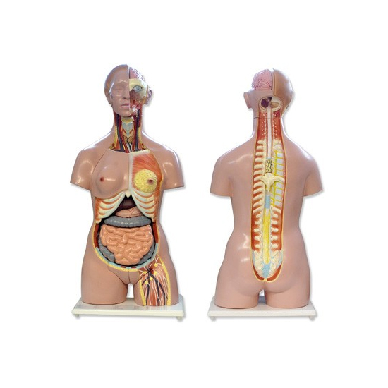
Dissectible in 12 parts, Male/Female reproductive organs can be easily taken out dissectible into 2 parts, without ribs cage.
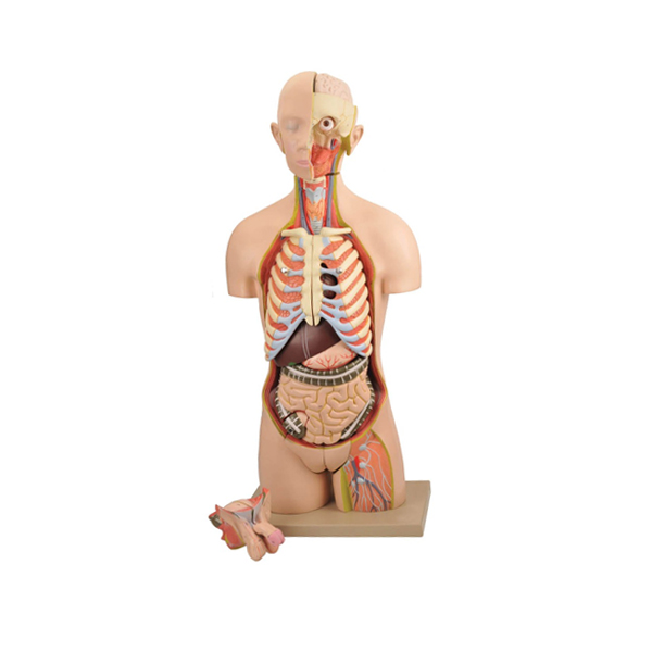
Dissectible in 12 parts, Male/Female reproductive organs can be easily taken out dissectible into 2 parts, without ribs cage, mounted on base with key card.
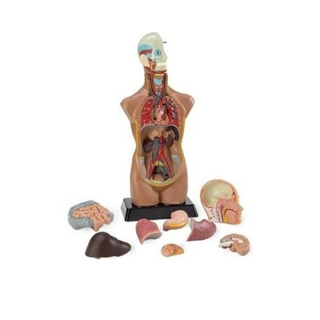
This life size torso composed of 12 parts, with interchangeable male and female genitalia, mounted on a base.
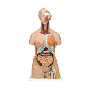
This life size torso composed of 24 parts, with interchangeable male and female genitalia, mounted on a base. All of the major organ systems are represented with great attention to accuracy and detail. Structures are numbered and identified on the accompanying key card.
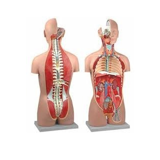
This sexless, life-size torso on base is accurate in all of its detailing. Anatomical structures of the major body systems are numbered an identified on the accompany key. The head is sectioned to expose one half of the brain. The neck is dissected through the ventral surface to show muscular, neural, vascular, and glandular structures. The thorax and abdomen are completely open, for an unrestrict.
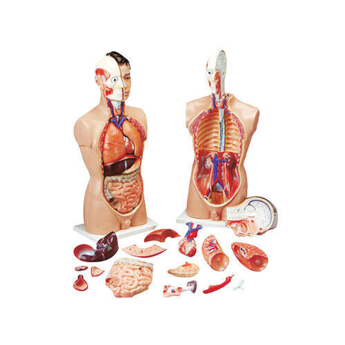
Dissectible in 17 parts, this life size torso with open back is provides an exceptionally realistic reproduction of anatomical structures in their finest detail.
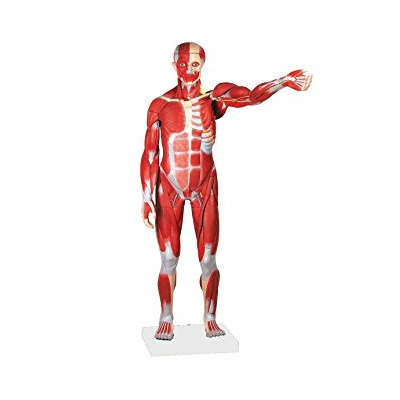
Complete human muscular and organs anatomy. It illustrates with excellent detail the superficial and deeper muscles, tendons, ligaments, vessels and body structures. The internal organs are removable for closer examination to study the relationships among all the body structures, mounted on base with stand.
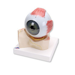
Dissectible into 6 parts, sectioned horizontally, upper half of the sclerotic membrane, choroids membrane, retina with vitreous humor, lens, lower half of the sclerotic membrane on base with key card.
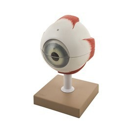
Eye model 5x enlarged, dissectible into 6 parts, showing both halves of sclera with cornea and eye muscle attachments and choroid with iris and retina, lens, vitreous humor mounted on a stand with numbered key card.
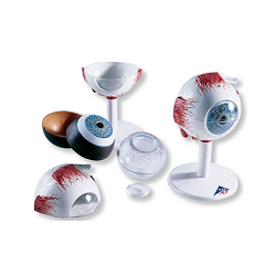
Dissectible into 6 parts, showing both halves of sclera with cornea and eye muscle attachments and choroid with iris and retina, lens, vitreous humor mounted on a stand with numbered key card.
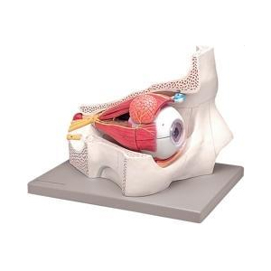
Dissectible into 7 parts, 4X life size, shows the eyeball with optic nerves and muscles in its natural position in the bony orbit, mounted on a stand with numbered key card.
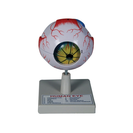
Dissectible into 8 parts, 5 Times enlarged model includes eyelids showing the bony orbit with intraocular muscles, a tear duct, and lachrymal gland. Cornea, muscles, 2 part choroid with iris, retina, and optic nerve; clear lens; and clear vitreous humor are removable, mounted on a stand with key card.
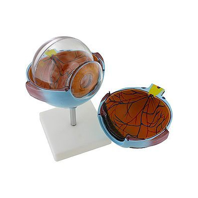
One side of this 6x model shows the eye socket with a sagittal cut away, while the other side of the model shows the outside of the eye and all the muscle attachments, as well as illustrations depicting how the eye focuses. The cut away is encased in a clear protective covering and two separate insets show the fine structure of the retina as seen with an electron microscope, on a base with Key Card.
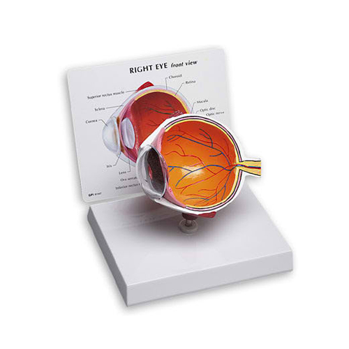
Enlarged approx. 6 times, 5 parts mounted board.
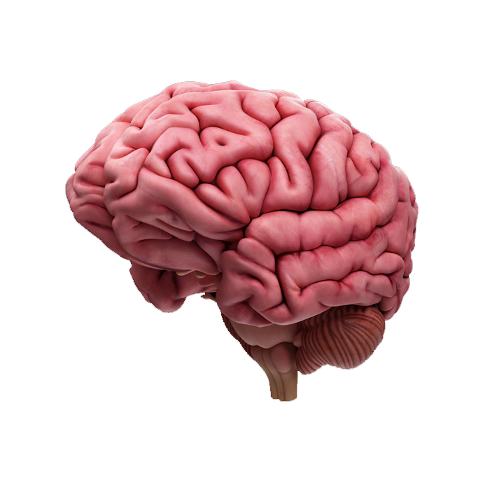
A life size brain model divided medially in 4 parts along the sagittal plane. It shows the left and right cerebrum, cerebellum, brain stem and blood vessels, with Key Card.
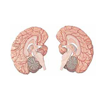
A life size brain model divided medially in 2 parts along the sagittal plane. It shows the left and right cerebrum, cerebellum, brain stem and blood vessels, with Key Card.
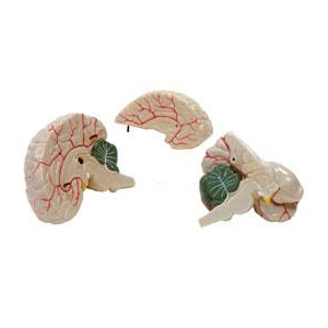
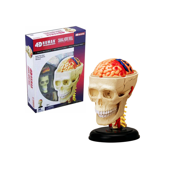
Dissectible in 3 parts, full size, showing important parts of skull and cranial nerves, mounted on base with key card.
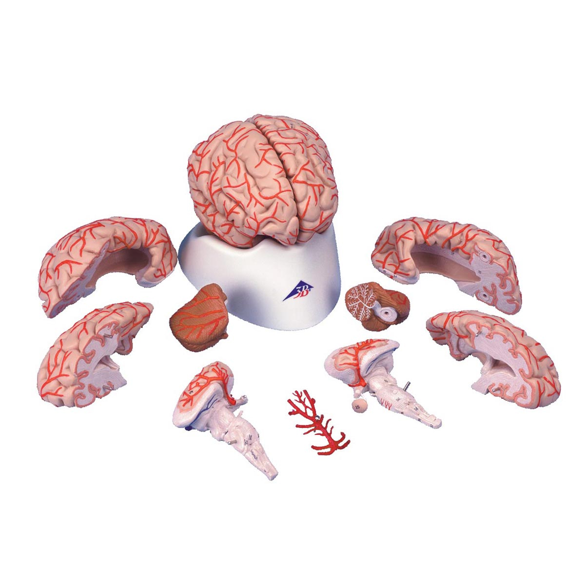
This brain model shows the brain arteries, separate altogether into 9 parts but mounted in normal position on a base with numbered Key Card.
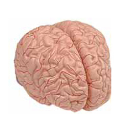
This brain model shows the brain arteries, separate altogether into 9 parts but mounted in normal position on a base with numbered Key Card.
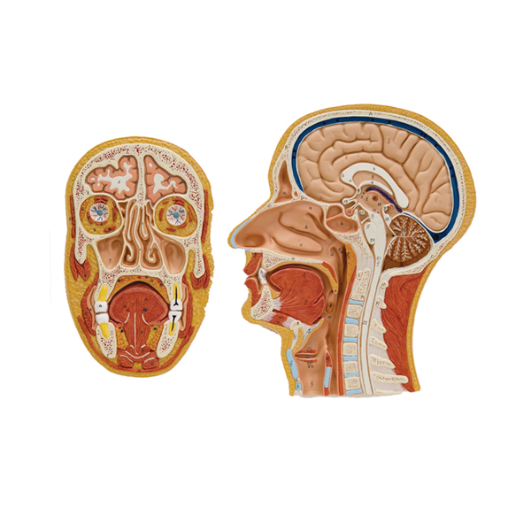
Life size representation of the superficial and the internal structures of the head and neck. Comparison between frontal and median sections provides a superior understanding of the anatomical structures of the head and neck, mounted on board with key card.
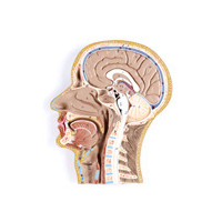
Life size representation of the superficial and the internal structures of the head and neck. This relief model shows all relevant structures of the human head in great detail. Mounted on board. With Key Card.

Enlarged approximately 2500 times, showing neuron structure clearly based on the latest electron microscopy. Showing medulated nerve fiber, axon, myelin sheath and nodes of ranvier etc., mounted on base with Key Card.
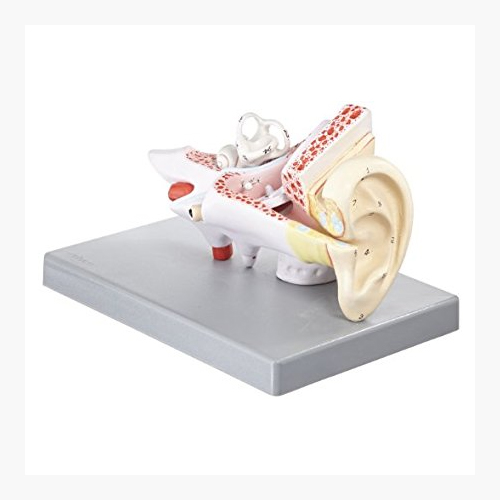
3X life size. The model shows the external, middle and inner ear. The eardrum with malleus, incus and stapes are removable. The other removable parts are composed of: cochlea and labyrinth with vestibular and cochlear nerves, 2 bone sections that define the middle and inner ear.
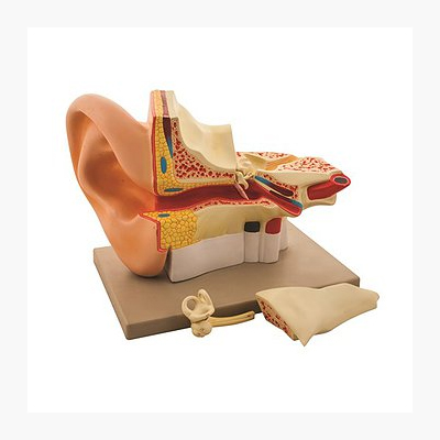
5 times life-size and in 3 parts, showing outer, middle and inner ear, removable auditory osicles and labyrinth with cochlea and vestibulocochlear nerve, mounted on base with key card.
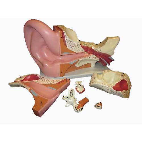
Dissectible into 3 parts, 4 times enlarged, showing petros portion of the temporal bone section of the auditory canal is removed, tympanic membrane with malleus and incus are also removable, mounted on base with key card.
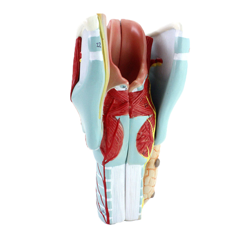
Dissectible in 2 parts, full size, showing muscles, thyroid glands and larynx, mounted on base with key card.
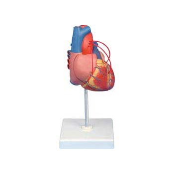
Life size in 2 parts, anterior heart wall can be removed to show the left and right ventricles and atria as well as the tricuspid, pulmonary, mitral and aortic valves, mounted on base with stand and key card.
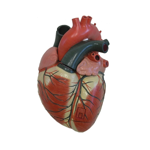
3 times life size and separates into four parts for detailed study. Sectioned so that both ventricles and atria open to expose the valves & large blood vessels near the heart and musculature of the heart are also shown, mounted on base with Key Card.
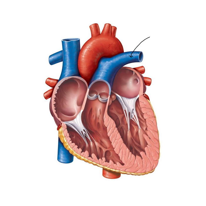
Dissectible into 2 parts, removable from base, shows internal and external anatomy including valves, cardiac chambers, and pulmonary and systemic vascular structures. Mounted on base, with key card.
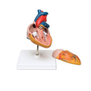
Human Heart Economy model dissectible in 2 parts, natural size, showing frontal section and the front of the heart on base, with key card.

Human Heart model dissectible in 3 parts, showing, Bicuspid and tricuspid semilunar and sigmoid values on base, with key card.
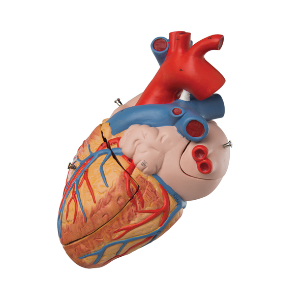
Dissectible in 4 parts, this model is enlarged 2 times the life size for a closer look at the intricate inner structure. The model is mounted on base with key card.
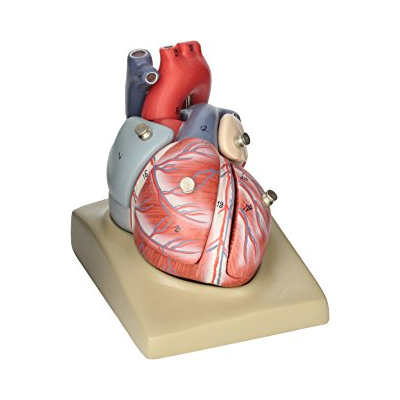
This model shows the anatomy of the human heart can be dissectible in 7 parts. The model is mounted on the base and includes a key Card for identifying parts: Oesophagus, Trachea, Superior vena cava, Aorta, Front Heart Wall, Upper half of the hearts.
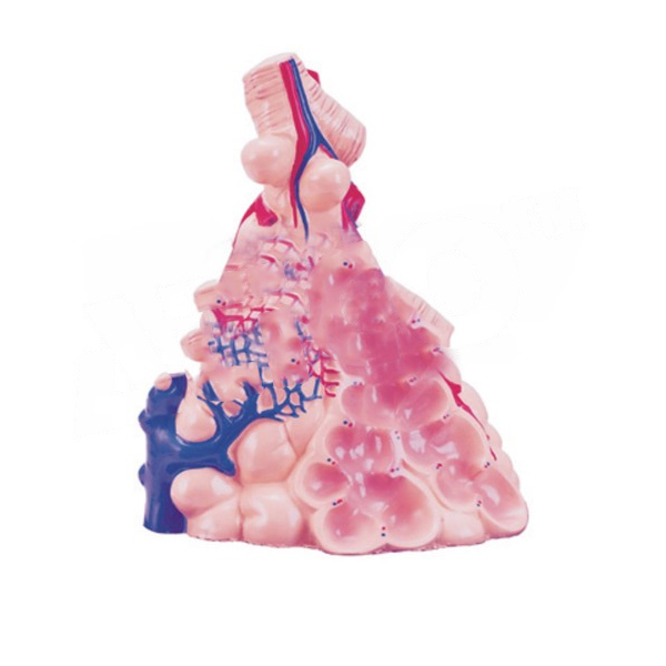
Model showing the structures involved in gaseous exchange, the alveolar epithelium, the alveoli, the capillary network, arteries, veins, bronchioles connecting to alveolar ducts, elastic fibers, smooth muscle, mucous membranes, and cartilage, mounted on a stand with key card.
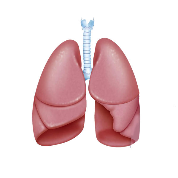
Dissectible in 2 parts, mounted on base with stand, showing all important parts with key card.
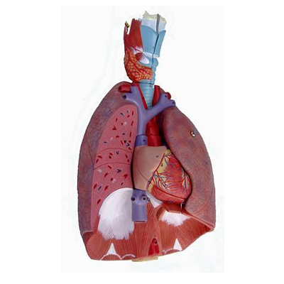
An actual size reproduction of the whole respiratory system, dissectible in 4 parts, showing the position of lungs diaphragm and heart, mounted on base with key card.
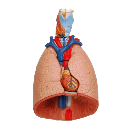
Dissectible in 2 parts, showing Larynx Trachea with bronchial tree, vena cava, aorta, 2-part heart, subclavian and vein, pulmonary artery, two parts lungs, diaphragm, oesophagus, mounted on base with key card.

2 Times Enlarged, dissectible in 5-part it is sectioned longitudinally to reveal intimate details of internal structures, including the hyoid bone, cartilages, ligaments, muscles, vessels, nerves and thyroid gland, mounted on base with key card.
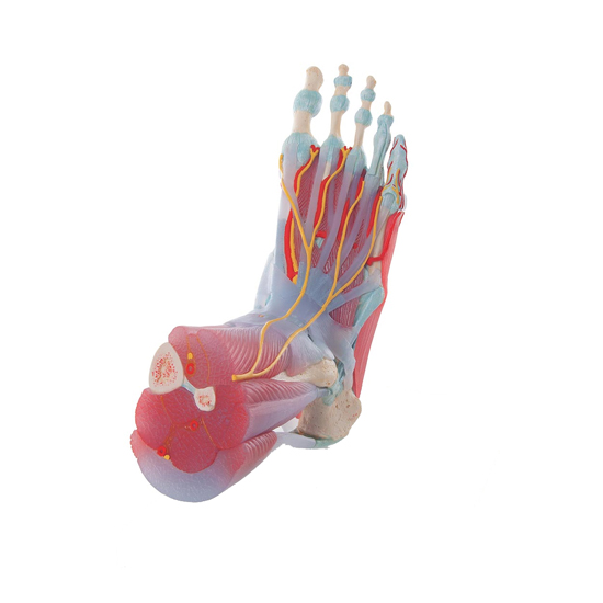
Life size model, dissectible in 3 parts. The plantar aponeurosis and the flexor brevis can be removed to show the underlying network of muscles, tendons, vessels and nerves. A deeper plantar dissection allows observation of the plantar muscles and plantar nerve branches. A superficial dorsal dissection shows ligaments, nerves and vessels.
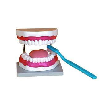
3x life size model, useful for teaching to brush teeth including giant toothbrush on base with key card.
2 parts, Lower Molar with root approximately 12X life size; it shows the most important dental pathologies, mounted on base with stand and key card.
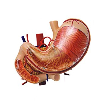
This life size model is dissected along the medial plane and can be opened to show the internal structure of the stomach, including the mucosa, the pylorus, and section of the gastric wall. The model also shows the superficial muscular layers with nerves and vascular structures and the blood vessels, mounted on base with stand and key card.
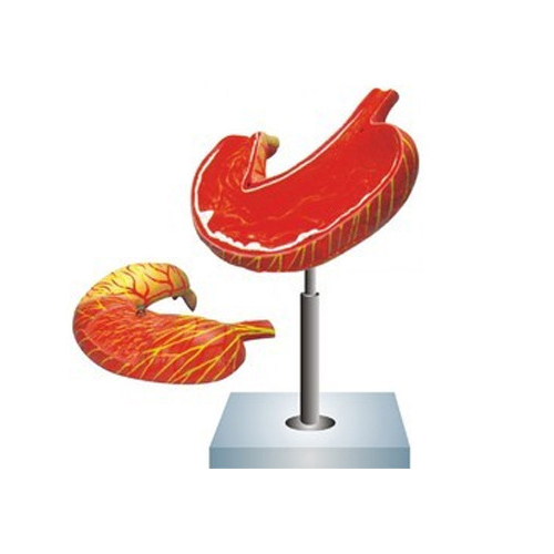
This life size model is dissected along the medial plane and can be opened to show the internal structure of the stomach, including the mucosa, the pylorus, and section of the gastric wall. The model also shows the superficial muscular layers with nerves and vascular structures and the blood vessels, mounted on base with stand and key card.
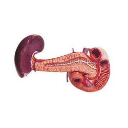
Life size model is an accurate representation of the pancreas, spleen and duodenum. The pancreas is open to show the entire pancreatic duct. The duodenum is partially dissected to expose its internal structure, mounted on base with stand.
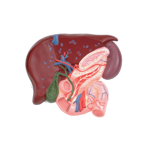
Model is 1.5 times enlarged, showing the branch of vessels in liver and the bile duct system, mounted on a base.
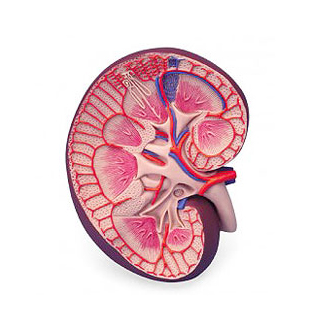
Life size model, 2 parts model of the kidney with the adrenal gland. The model is sectioned along the front plane; showing internal structures, including the cortex, medulla, pyramids with papillae, partially open renal pelvis, ureter, blood vessels.
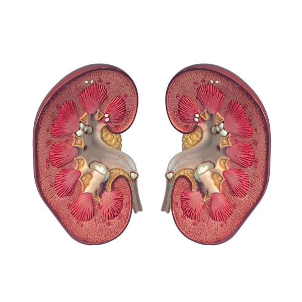
Showing longitudinal section of right kidney with its important parts, dissectible in 2 parts, enlarged about 3 times, on a base with key card.
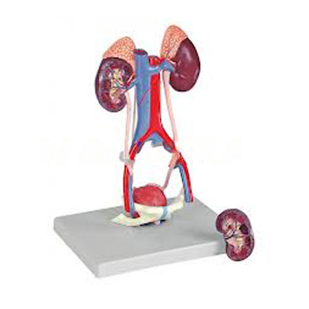
Showing the major components of the urinary system, plus the vena cava and abdominal aorta; the right kidney is dissected to show the cortex, medulla, pyramids, calyces, pelvis and origins of the renal artery and vein. The bladder can be opened to reveal the mucosa, trigone, urethra, seminal vesicles, ejaculatory ducts and vas deferens, on a base with key card.
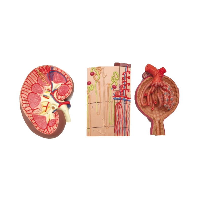
This 3 models set shows the basic structure of the kidney. The first model, a frontal section of the kidneyenlarged 3 times, illustrates the adrenal gland, cortex, medulla, pyramids with papillae, renal pelvis and blood vessels. The second model, representing a nephron enlarged 120 times, shows a renal tubules, a collecting tube system and Henle's loop. The third one illustrates Malpighian corpuscle with the Bowman's capsule, 700 times life size. All these models are a great aid to understanding the kidney anatomy in all its inner details, mounted on a base with key card.
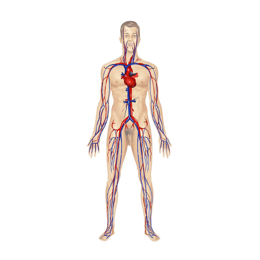
The model represents a general view of the human circulation. It includes the heart (2 parts), lungs, liver, spleen, kidneys and relevant connections with the pulmonary and systemic circulatory pathways.
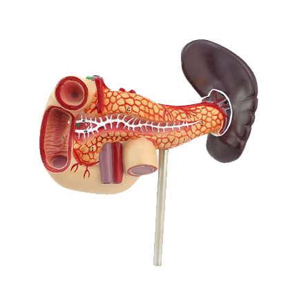
Model showing abdominal organs in detail. To understand the roles the liver, spleen, pancreas, and duodenum, process of digestion, life-size mounted on a stand with key card.
Life size, female pelvis showing female reproductive organs, bladder, uterus, rectum and ovary, mounted on base with key card.
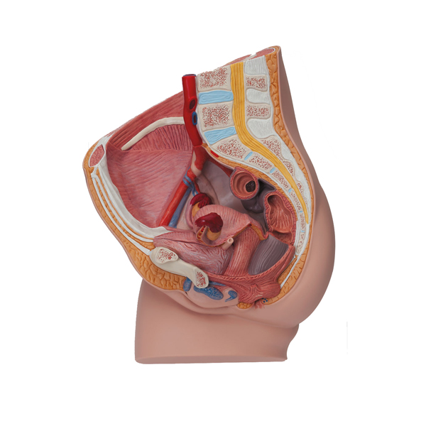
Life size, 2 parts showing median section, showing one half of female genital organs with bladder, rectum is removable, one half is shown at the normal position in the female pelvis, with key card.
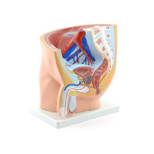
Life size, male pelvis showing internal and external reproductive organs and other vital organs of the pelvic area, mounted on base with key card.
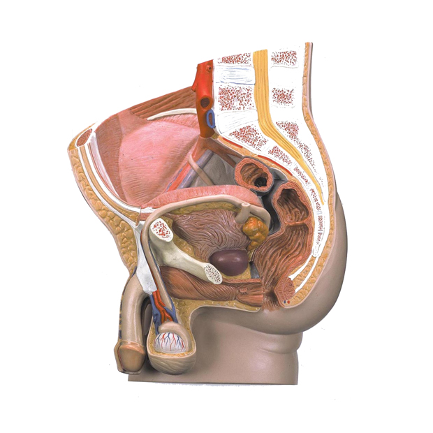
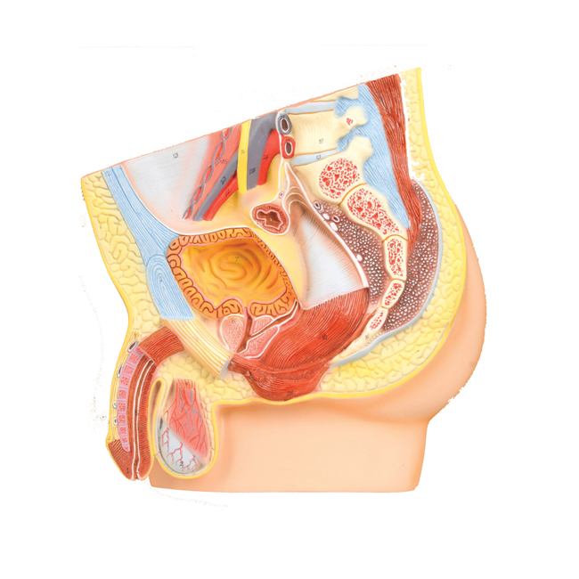
Life size, 2 parts showing median section showing one half of male genital organs with bladder, rectum is removable, one half is shown at the normal position in the male pelvis, with key card.
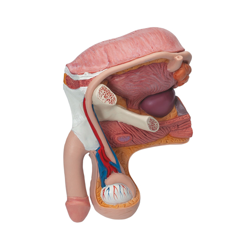
Natural Size, showing the internal and external genital organs of the pelvis, dissectible in 4 parts with key card.
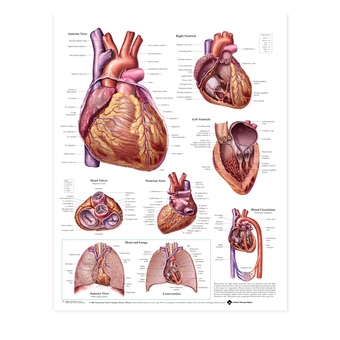
Stages of fetal development shown in this set of eight life-size models, each a re-creation of the uterus with a fetus. Models show a fetus at different times during the gestation period, mounted on individual stands with numbered key card.
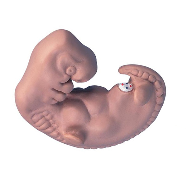
This model, 50X life size, shows structural details of a human embryo at 4 weeks of development.
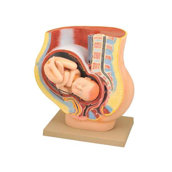
Life size model depicts the female human pelvis in median section with a fetus in the 40th week of pregnancy. The fetus is in the normal position prior to birth, showing the anatomical relationship between various structures of the maternal pelvis and the fetus. Fetus is detachable for closer examination.
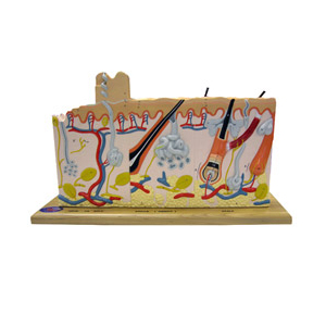
Model showing a section through the three layers of the hair covered skin of the head, representation of hair follicles with sebaceous glands, sweat glands, receptors, nerves and vessels.
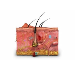
70 times enlarged, this model shows the structure of the human skin in 3 dimensions.
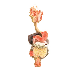
This life size model shows the human digestive tract from mouth cavity to rectum. The oral cavity, the pharynx and the first part of the oesophagus are dissected along the medial sagittal plane. The liver is shown together with the gall bladder and the pancreas is dissected to expose internal features. The stomach is open along the frontal plane; the duodenum, caecum, part of the small intestine and the rectum are open to expose the interior. The transverse colon is removable.
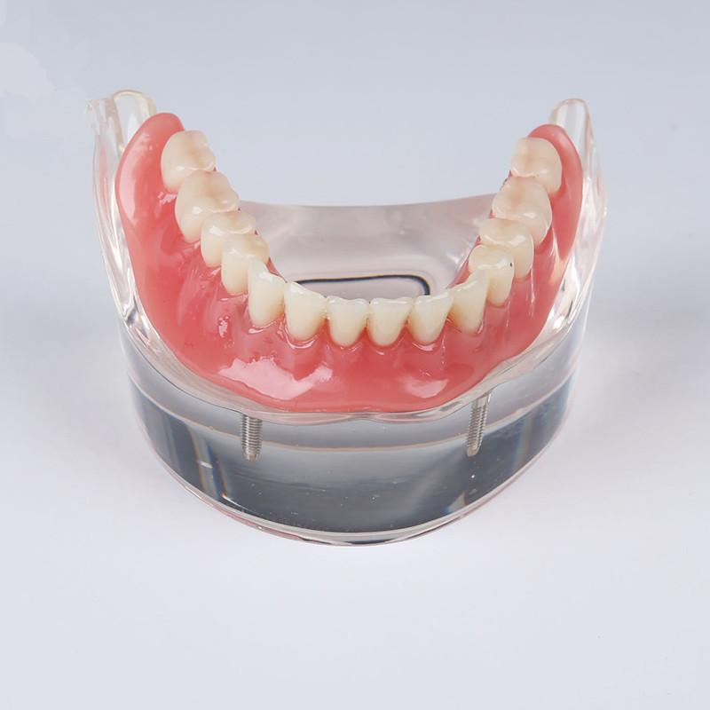
Greatly enlarged, for showing all the position of tooth the inner wall is removed, mounted on board with Key Card.
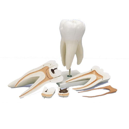
Human Teeth 6 Parts with Molar Anatomy Model - Greatly enlarged, for showing all the position of tooth along with upper triple root molar and caries, 6 parts, mounted on base with Key Card.
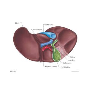
Human Liver Anatomy Model - Model is 1.5 times enlarged, showing the branch of vessels in liver, mounted on a base, with Key Card.
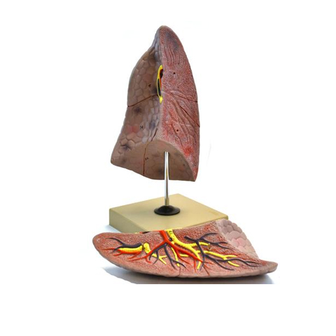
Human Lungs 4 Parts Anatomy Model - Dissectible in 4 parts, mounted on base with stand, showing all important parts with key card.
Life size model is an accurate representation of the spleen and duodenum, mounted on base.
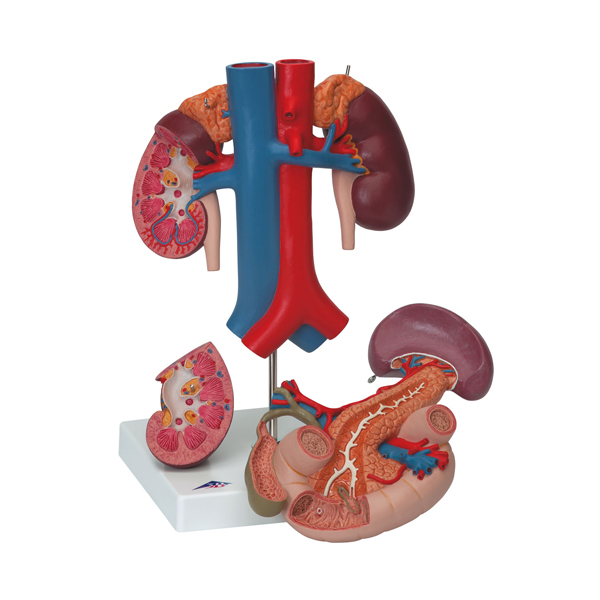
Model is an accurate representation of traverse section of human abdomen at level of omentum foramen, mounted on base.
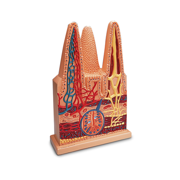
Model is an accurate representation of entire Intestinal Villus show arterioles and venules , mounted on base.
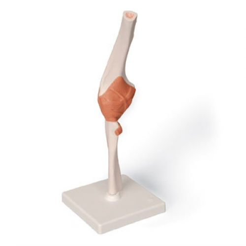
Model of human elbow joint , mounted on base.
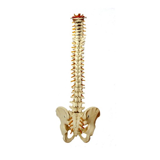
Model of human long backbone and oslamina , mounted on base.
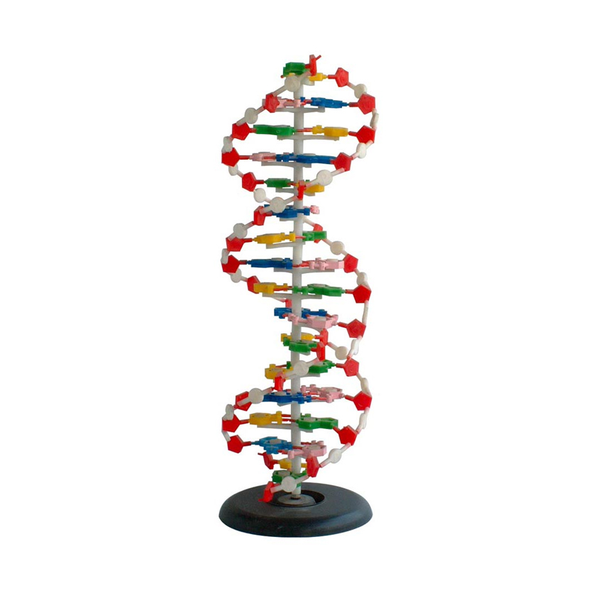
Model of human D.N.A showing its segments molecule mechanism of replication, structural formula, molecule shape and bone & angles distorted, mounted on board.
Model of human D.N.A, 3-Dimensional.
Human Nasal Cavity Model - This model shows section of the nasal cavity. On stand. PVC Human Nasal Cavity Anatomy Model.
Ambala Science Market © all rights reserved